
Cronulla Veterinary Clinic treats eye problems in pets. Servicing Cronulla, Caringbah and Sutherland Shire.
Eye problems in pets are a common reason to visit the vet. Our pets can suffer from the same eye problems as people, including cataracts, glaucoma and corneal injuries. Before you can figure out which problem your pet may have, you should learn a bit about how to eye works and how these diseases manifest.
Common Eye Problems
The eye is made up of three layers. The outer layer is made up of the clear cornea at the front and the white fibrous protective sclera around it. The inner layer is the light sensitive retina at the back of the eye. The layer in between is the uvea, rich in blood vessels, made up of the iris at the front, the ciliary body (that produces fluid inside the eye) and the choroid at the back which feeds the retina.
The three main disease processes that cause “red eye” are:
- Conjunctivitis
- Uveitis
- Glaucoma
As a precaution, if your pet’s eye or eyes look red, it is important to visit your vet as soon as possible.
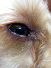
Conjunctivitis
Conjunctivitis literally means inflammation of the conjunctiva, the structures surrounding the eye.
As an illness, conjunctivitis can be acute or more chronic, and can be caused by many things such as eyelid abnormalities, infections, “dry eye” (insufficient tear production), mechanical or chemical irritations (such as an eyelid growth or shampoo), allergies or even cancers.
Conjunctivitis can cause symptoms such as redness, puffy eyelids, watery eyes or colored mucous discharge, itchy eyes, and squinting. The symptoms can range from mild to severe. Sometimes conjunctivitis can even lead to complications such as dry eye and corneal damage.
Some of the above symptoms can also be seen with uveitis and glaucoma. However, these latter issues are much more serious conditions which could lead to blindness!
 Uveitis
 Uveitis
Uveitis literally means inflammation of the uvea (the middle layer of the eye, rich in blood vessels, which is the “nutritional” layer of the eye).
Uveitis can have many causes, and sometimes no cause can be found.
Local causes can include trauma to the eye (such as the eye being hit by a golf ball, or a corneal ulcer), lens damage such as cataracts, and tumours.
Systemic causes can include certain infections (FIV, FeLV, Feline flu, distemper, toxoplasmosis), immune mediated diseases, high blood pressure, Diabetes Mellitus, and several other conditions.
Acute uveitis is a painful condition. We can tell the eye is painful because of squinting, light sensitivity, fluid discharge, and (in species which have it) the 3rd eyelid covering part of the eye. Looking closely at the eye will reveal that the sclera and conjunctiva are red and the cornea can be cloudy and hazy (due to corneal oedema) with a narrow pupil. If left untreated, the inflammation can cause vision loss and the eye can develop lens cataracts or secondary glaucoma. In chronic cases the eye may also lose internal eye pressure and shrink.
Uveitis needs aggressive treatments with regular follow ups to reduce the risk of complications and vision loss.
Glaucoma
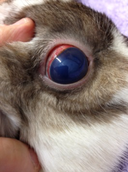 Glaucoma is an increase in the pressure within the eye. Fluids are constantly produced and drained within the eye. These fluids nourish the eye and maintain its shape with a normal intraocular pressure of 15 to 20 mm Hg.
In cases of glaucoma, something often goes wrong with the drainage of fluids from the eye, causing a buildup of pressure. This will cause a reddened eye, a hazy blue cornea, possible fluid or pus discharge, a large pupil, and pain which may show as a head shyness, hiding, or irritability. Later on the eye can become larger in size. Untreated, glaucoma leads to blindness by permanently damaging the optic nerve in the back of the eye.
Glaucoma can be primary (inherited, breed specific) or secondary (as a result of a separate underlying problem in the eye).
Primary glaucoma is seen in dog breeds such as the American Cocker Spaniel, Basset Hound, Chow Chow, Shar Pei, Jack Russell Terrier, Shih Tzu, and Arctic Circle breeds, but is rare in cats. Primary glaucoma usually begins in one eye, but eventually affects both eyes in most patients. This makes early diagnosis very important!!
Secondary glaucoma can develop whenever a condition exists that interferes with the natural flow of the ocular fluid. Examples of conditions that can lead to secondary glaucoma are uveitis, lens dislocation, tumours in the eye, and eye injuries.
When diagnosing eye problems, we use different tests and techniques to come to a diagnosis, which informs decisions about the best treatment options.
We will routinely start with a standard patient history and physical examination. The eye examination consists of visual examination of the eyes and structures around the eyes, as well as an examination of the inner parts of the eyes with an ophthalmoscope. Tear production may be checked with a Schirmer Tear Test, fluorescein drops may be applied to the eye, and other drops may be applied for further investigations. In some cases further tests including blood and urine tests may be needed, and complicated cases may need to be referred to an eye specialist.
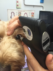 To check the internal pressure of your pet’s eyes, our clinic uses a Tonovet Tonometer, the latest in intraocular pressure diagnostics. This procedure has many advantages; for example, there is no need for sedation or eye drops, it is easy and quick to use, and it causes no discomfort to your pet. This tool can be used for diagnosis of eye pressure problems and monitoring the effectiveness of treatments.
For further information on eyes problems in pets, please contact our clinic to speak to our friendly staff or book an appointment.